Medical Tech Outlook Weekly Brief
Be first to read the latest tech news, Industry Leader's Insights, and CIO interviews of medium and large enterprises exclusively from Medical Tech Outlook
A featured contribution from Leadership Perspectives: a curated forum reserved for leaders nominated by our subscribers and vetted by our MedTech Outlook Advisory Board.


Following the new ESC guidelines it seems extremely important the key role of “Heart Team” for planning the perfect surgical approach. The Heart Team, in selected cases, in patients that cannotbear the open surgery,suggest the percutaneous mini-invasive approach as a good compromise for aortic and mitral valve repairment.
Nowadays the pre-procedural planning of percutaneous mini-invasive surgery is strictly dependent by the acquisition of CT and MR.
Both these techniques provide a comprehensive evaluation of valve anatomy, coronary arteries and vascular access; these CT findings provide a pivotal rule for the correct planning of surgery.
In patients that should undergo to transcatheter aortic valve replacement (TAVR) the role of CT is fundamental forthe evaluation of aortic anulus, calcification of the aortic valve, coronary arteries and vascular access. The evaluation of aortic anulus can provide pivotal information regarding the sizing of the prosthetic aortic valve furthermore the calcification of the valve can have an important impact for expansion of the implanted valve. The evaluation of coronary artery in patients with TAVR can be extremely importantin order to avoid the unnecessary invasive coronary angiography (ICA), considering the powerful rule-out ability of cardiac computed tomography angiography (CCTA) for presence of coronary artery disease. Furthermore, in patients candidated for TAVR the simultaneous acquisition of CT angiography during the acquisition of CCTAcan be extremely important for the evaluation of arterial access for the presence of kinking, coiling or narrowing due to atherosclerotic plaques.
Regarding the transcatheter mitral valve repairment (TMVR)the CT is a fundamental tool for the evaluation of mitral valve anulus and cardiac surrounding structures.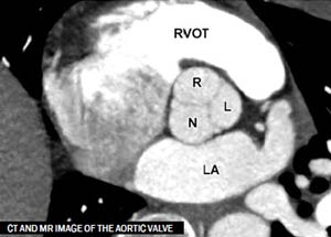
If the patient is selected for percutaneous mitral annuloplasty beyond the dimensions of mitral anulus,CT can be also important for the assessment of surrounding cardiac structure. In particular the following CT finding needs to be carefully evaluated: mid anchor to LV distance >4mm, mitral leaflet hinge point <8mm, anchor-Left circumflex > 2.5mm, relationship with coronary sinus. It is fundamental to consider the possibility to evaluate with CT, in the same acquisition of the heart the systemic venous system; the latter will be extremely helpful for the planning of the better vascular access. Furthermore in case of mitral valve replacement the CT study can be extremely helpful for the evaluation of new left ventricle outflow tract in order to exclude the obstruction of the outflow with new valve.
“The evaluation of aortic and mitral anulus with CT can provide pivotal information regarding the sizing of the prosthetic valve furthermore the relationship with surrounding structures are important in order to avoid surgical complication”
Cardiac magnetic resonance represents an alternative techniques that allows the evaluation of valve anatomy in both TAVR and TMVR with a better temporal resolution compared to CCTA without use of radiation dose. Also with Magnetic resonance it is possible to evaluate vascular access, however compared to CT it is extremely difficult to evaluate the presence of calcified atherosclerotic plaques.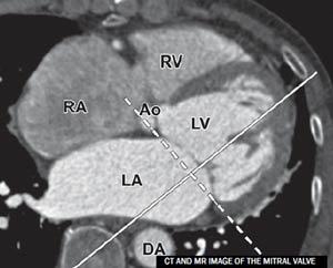
Considering the future and important application of percutaneous transcatheter repairment, the technology is rapidly moving to techniques that can provide information useful for mini-invasive surgical approach preferably with low dose or absent contrast agent in both CT and MR. In the field of CT,considering that the patients candidate to mini-invasive approach are old patients, sometimes with impairment of renal function the CT protocols, as described above, are focused on the reduction of iodine contrast agent and low-radiation dose. While in MR is still mainly used in patients that cannot undergo to CT.
Furthermore, following the ESC guidelines,the non-invasive evaluation of patients with valvular heart disease will be only a step for a complete evaluation of the Heart Team.